Hartlepool & Stockton Health

  • Our Services
    • SPLW
  • About us
    • Our Story
    • CQC
    • Our Plans
  • Our people
    • Join our Team
  • Latest news
    • Casebooks
    • Newsletter
  • Contact us
    • Privacy Notice
  • H&SH updates
  • Stakeholder area
  • Resources
  • COVID-19
    • Tees Valley COVID-19 Virtual Ward

Casebooks

Lucy’s case book issue 3 - the ruptured aortic aneurysm

4/11/2019

0 Comments

 
Overview
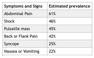
The ruptured Abdominal Aortic Aneurysm is a medical emergency which is often fatal. The classic triad of “shock, abdominal pain and a pulsatile mass” is not always present and the urgency of the situation not therefore recognised1. It is suggested that a misdiagnosis rate for ruptured Aortic Aneurysms may be as high as 42% with incorrect diagnosis including renal colic (6%), myocardial infarction (6%), colonic inflammation (3%) and gastrointestinal perforation (3%).

​This issue persists and has not been improved by better diagnostics and surgical imaging. As the frequency of Aortic Aneurysms and their acute rupture diminish with screening and falling cardiovascular disease rates the diagnostic challenge for a rare event will become harder.

Signs and Symptoms
Picture

The rupture of an aortic aneurysm may occur abruptly and catastrophically or can evolve over a few hours or days as the leak is tamponaded by surrounding tissues. 
​
​
The symptoms that occur depend upon the location of the bleeding, with ~80% of ruptures being retroperitonieal2.

The conditions prevalence increases with age over 55, smoking and hypertensive history with males developing the condition 10 years earlier than women3,4 Clearly if the patient is known in advance of symptoms to have an enlarged aorta then raising concern regarding a possible diagnosis in the presence of back or abdominal pain is somewhat easier.

The rare and unusual presentations can include


  • Transient lower limb paralysis
  • Right hypochondrial pain
  • Nephroureterolithiasis
  • Groin pain
  • Testicular pain
  • Testicular ecchymosis (blue scrotum sign of Bryant)
  • Iliofemoral venous thrombosis
  • Inguinoscrotal mass mimicking a hernia

Actions
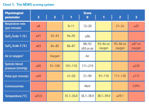
Ruptured aortic aneurysm should not be discounted from the differential diagnosis of sudden back or abdominal pain, because hypotension or a pulsatile mass are absent. Consideration should be given to taking physical observations and calculating a NEWS score where there is diagnostic uncertainty. Awareness of some of the other features which may present is likely to be helpful.

Summary
  • Ruptured aortic aneurysm is a fatal surgical emergency
  • The classic triad of pain, hypotension and a pulsatile mass is present in only 25-50% of patients
  • Different sites of aneurysm rupture determine the various clinical presentations seen in patients
  • Early recognition of these presentations and prompt treatment of ruptured aneurysms is life saving

1.    Azhar B, Patel SR, Holt PJE, Hinchliffe RJ, Thompson MM, Karthikesalingam A. Misdiagnosis of Ruptured Abdominal Aortic Aneurysm: Systematic Review and Meta-Analysis. Journal of Endovascular Therapy 2014; 21(4): 568-75.
2.    Assar AN, Zarins CK. Ruptured abdominal aortic aneurysm: a surgical emergency with many clinical presentations. Postgrad Med J 2009; 85(1003): 268-73.
3.    Reite A, Soreide K, Ellingsen CL, Kvaloy JT, Vetrhus M. Epidemiology of ruptured abdominal aortic aneurysms in a well-defined Norwegian population with trends in incidence, intervention rate, and mortality. Journal of vascular surgery 2015; 61(5): 1168-74.
4.    Aggarwal S, Qamar A, Sharma V, Sharma A. Abdominal aortic aneurysm: A comprehensive review. Experimental and clinical cardiology 2011; 16(1): 11-5.

Download the PDF.
0 Comments

Lucy’s case book issue 2 - sepsis and septic shock.

4/11/2019

0 Comments

 
Overview
Sepsis is responsible for ~37,000 deaths a year in England, with 123,000 cases annually1
. Sepsis can be difficult to spot and good GPs “Miss It”. Sepsis is defined as “life threatening organ dysfunction caused by a dysregulated host response to an infection2” and is a time critical medical emergency. The mortality of Septic Shock increases 8% with every hour that passes without initiation of iv antibiotics3. If recognised early it can be treated effectively with oxygen, fluids, and antibiotics4.

Signs and Symptoms
Infection is a common cause of ill health but there are few pathognomonic signs or symptoms specific to deteriorating infection or sepsis. NICE list symptoms that may alert to the possibility but over triage significantly in most locations.( Appendix A).


Assessing Adults
The presence of sepsis is most commonly indicated by a history of deterioration in the presence of possible infection, clinical judgement and abnormal physiology. The assessment of physiology should include respiratory rate, blood pressure/perfusion, oximetry, pulse and temperature, with a judgement made about cognition.  We know that the presence of abnormal respiratory rate, reduced blood pressure and altered cognition are the best predictors of sepsis particularly in combination. In hospital abnormal values for these variables are assessed against a NEWS Score using the table below to calculate an aggregated score. The predictive value of NEWS in Primary care is yet to be determined, but we know that patients presenting in the ED with a NEWS score of 3 or less have a low chance of having sepsis, and that a score of 5 or above is strongly suggestive of sepsis in the presence of probable infection.


Picture
For GPs this amounts to  identifying  a deteriorating patient with possible/probable infection  using clinical judgement and augmenting that decision making by assessing physiology. When a decision is made to admit this should be done by the clinician calling 999, stating that they “Suspect Sepsis” and being prepared to give the NEWS Score or the abnormal physiology of concern.​

In patients where sepsis is not suspected but infection is a possible or probable cause of being unwell they should have some recorded assessment of respiratory rate/effort, perfusion, and cognition. They or their carers should also receive specific safety netting as to the signs of sepsis, and ideally an age appropriate Sepsis leaflet.

Assessing Children
This can present a significant clinical challenge. Children’s physiology can vary wildly according to age and their response to infection. There is no standardisation of Paediatric Early Warning Scores as yet and the values they contain are validated against different cohorts of children to those seen in Primary care. There is also no standard paediatric definition of what determines the presence or absence of sepsis.

None of this helps the General Practice clinician. Clinical assessment should include judgement as to the history and unusual signs, including rashes abnormal alertness, clinician and parental concern. It should also contain a record of an assessment of physiology, particularly their respiratory rate/distress, perfusion (capillary refill, pulse rate) and level of cognition/social interaction.  In many children this may be represented as negative findings, (no respiratory distress, pink and playing with toys, running around room or smiling appropriately). Temperature alone is a poor predictor of wellness. The more unwell the child the more formal the assessment may need to be, particularly where the judgement is to keep the child at home.

Safety netting for children needs to be better than “if they get worse come back” and should include some indication of what worse looks like. This should be supported by an age appropriate leaflet.

Verbal advice for Parents of Children with Infection
Your child has xxx infection and is likely to improve with/without treatment. Very occasionally they can get significantly worse, signs of getting worse can include:


  • Becoming increasingly breathless 
  • Pale and clammy “the colour someone goes when they are about to be sick”
  • Unnaturally drowsy or unresponsive
  • Not passing urine in the previous 12 hours.

If these occur then you should consider getting your child assessed as a matter of urgency.

Summary

  • When assessing any patient with infection consider if there are features that might suggest sepsis and document their presence or absence
  • Assessment of Respiratory Rate, Blood Pressure/perfusion and cognition should all be assessed and recorded for all patients with infection
  • National Early Warning Score (2017) should be considered to be the bench mark for abnormal values in adults.
  • Paediatric Early Warning scores and NICE Guidance vary in their contents and values but all provide an indication of abnormal values. Clinical and parental judgement remains a strong predictors
  • Oximetry should be recorded using age appropriate devices and probes
  • Escalation to ambulance or other services should be done by the clinician and must include the phrase “Suspected Sepsis” and give a NEWS score in adults and abnormal physiological findings in children
  • Safety Netting should be sepsis specific supported by age appropriate leaflets.

Further Reading
E-Learning for Health Sepsis Modules
RCGP Sepsis: Guidance for GPs


References
1.    Hospital Episode Statistics: Health and Social Care Information Centre. 2015. http://www.parliament.uk/business/publications/written-questions-answers-statements/written-question/Commons/2015-09-16/10526/.
2.    Singer M, Deutschman CS, Seymour CW, et al. The Third International Consensus Definitions for Sepsis and Septic Shock (Sepsis-3). Jama 2016; 315(8): 801-10.
3.    Kumar A, Roberts D, Wood KE, et al. Duration of hypotension before initiation of effective antimicrobial therapy is the critical determinant of survival in human septic shock. Critical care medicine 2006; 34(6): 1589-96.
4.    Daniels R. Surviving the first hours in sepsis: getting the basics right (an intensivist's perspective). The Journal of antimicrobial chemotherapy 2011; 66 Suppl 2: ii11-23.

Appendix A
NICE Guidance  
Table 1: Risk stratification tools for adults, children and young people aged 12 years and over with suspected sepsis
Picture
Table 2: Risk stratification tool for children aged 5-11 years with suspected sepsis
Picture
Table 3: Risk stratification tool for children aged under 5 years with suspected sepsis
Picture

​Additional Resources
http://www.rcgp.org.uk/clinical-and-research/toolkits/sepsis-toolkit.aspx

​Download the PDF.
0 Comments

Lucy’s case book Issue 1 - testicular torsion (the painful testicle)

4/11/2019

0 Comments

 
Overview
Testicular torsion is a relatively uncommon presentation within regular general practice but has a suggested prevalence of 1 in 4,000 in males under the age of 25. It appears with greater frequency within providers of unscheduled care because of its relatively sudden and severity of presentation. It can be difficult to recognise clinically and can be intermittent in its presentation as the testes twist and spontaneously untwist. Once recognised it is a surgical emergency with a 6 hour window for testes preserving surgery.

Signs and Symptoms
These are variable testicular pain is not a universal finding and can be absent or intermittent. Swelling of the scrotum or testes, oedema and redness of scrotal skin or abdominal pain may all be features. It is important that the scrotum is carefully examined in the distressed male infant where no other cause is identified.

Typically the pain should be severe and sudden onset, possibly radiating to the groin or lower abdomen, it can be accompanied by vomiting nausea and dysuria. It may be proceeded by low grade trauma to the testes and should be considered where there is no history of urinary tract or Genito urinary infection

Picture

​Signs are of limited value in excluding the diagnosis, though the presence of a transverse high lying testes or abnormal position compared with the alternate side may support clinical suspicion.


Actions
Testicular torsion is a surgical emergency with testicular salvage possible in 90-100% of cases operated on within 6 hours of onset of symptoms, this drops to 20-50% at 12 hours after onset. Orchidectomy or subsequent testicular atrophy results in 5-50%
​
Patients should be referred for urgent assessment and kept nil by mouth until a torsion has been excluded in hospital.

Summary
  • Testicular torsion may be difficult to diagnose if symptoms are intermittent or atypical, but it must be considered in all cases of scrotal pain, with careful history and examination.
  • Sudden, severe onset of testicular pain with tenderness should be considered as torsion and referred, unless other clinical features suggest an alternative diagnosis.
  • Examine the testis for tenderness, size, shape, and position, and examine the remaining scrotal contents, comparing findings with the unaffected side.

Case Study
https://www.themdu.com/guidance-and-advice/case-studies/testicular-torsion

Learning resources.
https://bestpractice.bmj.com/topics/en-gb/506 
https://learning.bmj.com/learning/module-intro/testicular-torsion.html?moduleId=10029430&searchTerm=“testicular torsion”&page=1&locale=en_GB 

Further Reading
Testicular Torsion  
1.    Somani BK, Watson G, Townell N. Testicular torsion. BMJ (Clinical research ed) 2010; 341.

​Download the PDF.
0 Comments

    Author

    Write something about yourself. No need to be fancy, just an overview.

    Archives

    April 2019

    Categories

    All

    RSS Feed

Picture
21 Gloucester House, Church Road, Stockton-on-Tees  TS18 1TW
​
t. 01642 061047  |  e. info.hash@nhs.net

Website by Get Noticed Branding